Treatments
- Treatments
- Diagnostic Angiograms & Venograms
- Angioplasty & Stent Placement
- Aortic & Peripheral Stent-Grafts
- Transcatheter Embolization
- Transjugular Liver Biopsy
- TIPS (Transjugular Intrahepatic Porto-Systemic Stent Shunt)
- Intra-arterial & Venous Thrombolysis
- Inferior Vena Cava (IVC) Filter Placement
- Biliary Drainage Procedures
- Nephrostomy Tube & Nephroureteral Stent Placement
- Fallopian Tube Recanalization
- Musculoskeletal Embolization (Knee OA, Plantar Fasciitis)
- Prostatic Artery Embolisation
Peripheral & Visceral Pseudoaneurysm Embolization
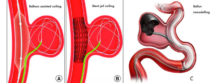
Peripheral and visceral pseudoaneurysms are abnormal dilations of arteries, often caused by trauma, infection, iatrogenic injury, or underlying vascular disease. Unlike true aneurysms, pseudoaneurysms involve a breach in the arterial wall with blood contained by surrounding tissue. Embolization is a minimally invasive, image-guided procedure used to treat these lesions, especially when surgical intervention is high-risk or not feasible. Using angiography, interventional radiologists access the affected artery via a catheter and deliver embolic agents such as coils, glue, or particles to occlude the pseudoaneurysm. This procedure effectively prevents rupture, controls hemorrhage, and preserves surrounding organ function. It has high success rates with low complication risks and allows rapid recovery compared to open surgery.
- Patient Evaluation: Assess pseudoaneurysm location, size, etiology, and patient comorbidities via imaging (CT angiography, Doppler ultrasound, or MRI).
- Access Site Selection: Usually via femoral or radial artery puncture under local anesthesia.
- Angiographic Mapping: A catheter is navigated to the artery supplying the pseudoaneurysm, and contrast dye is injected to delineate anatomy.
- Embolic Material Selection: Based on vessel size, flow dynamics, and pseudoaneurysm morphology; commonly used agents include coils, glue (NBCA), polyvinyl alcohol particles, or vascular plugs.
- Embolization: The embolic agent is deployed to occlude the pseudoaneurysm sac and/or feeding artery, ensuring complete cessation of blood flow into the lesion.
- Recovery & Follow-Up: Patients are monitored for complications such as pain, ischemia, or infection. Follow-up imaging ensures the pseudoaneurysm remains obliterated.